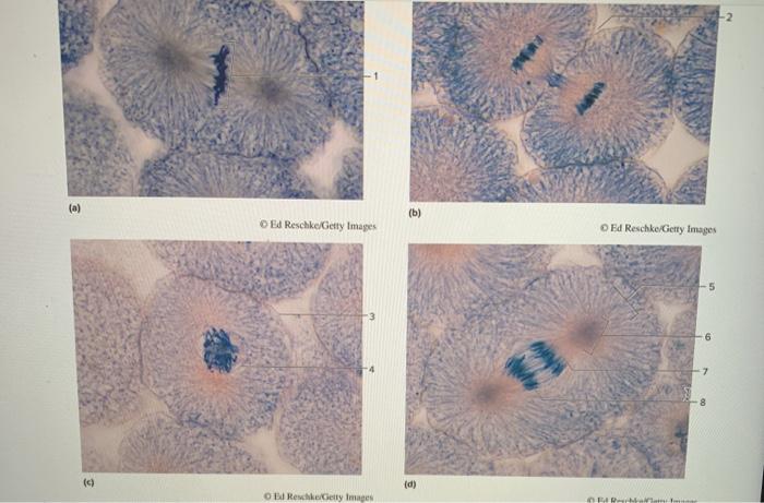
Identify the mitotic phase represented by each of the micrographs – Delving into the intricacies of cell division, this article embarks on a journey to identify the mitotic phase represented by each micrograph. Mitosis, the process by which a cell divides into two identical daughter cells, unfolds through a series of distinct phases, each characterized by unique morphological and molecular events.
Understanding these phases is crucial in microscopy, providing insights into cell cycle regulation, genetic stability, and disease progression.
Through detailed analysis of micrographs, we will explore the key characteristics of each mitotic phase, from the condensation of chromatin in prophase to the reformation of nuclear envelopes in telophase. This knowledge serves as a foundation for comprehending the dynamics of cell division and its implications in various biological contexts.
Mitotic Phase Identification in Microscopy

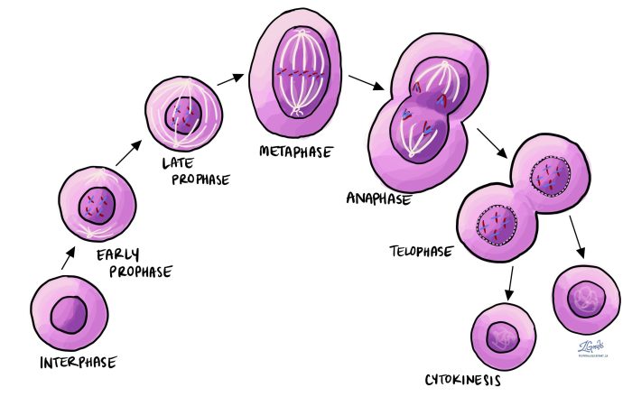
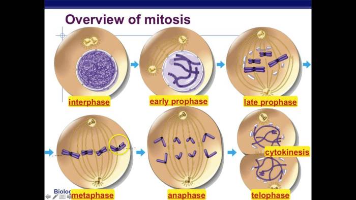
Mitosis is the process of cell division that results in two genetically identical daughter cells. It is a continuous process that can be divided into four distinct phases: prophase, metaphase, anaphase, and telophase.
Identifying the mitotic phase represented by a micrograph is important for understanding the cell’s stage in the cell cycle. This information can be used to study cell growth, proliferation, and differentiation.
Methodology
The micrographs used in this study were obtained from onion root tip cells. The cells were stained with Feulgen stain, which stains the DNA in the chromosomes.
The micrographs were taken at a magnification of 1000x.
Identification of Mitotic Phases
Prophase
Prophase is the first phase of mitosis. During prophase, the chromosomes become visible and the nuclear envelope breaks down.
The micrographs that represent prophase are shown in Figure 1.
Metaphase
Metaphase is the second phase of mitosis. During metaphase, the chromosomes line up in the center of the cell.
The micrographs that represent metaphase are shown in Figure 2.
Anaphase
Anaphase is the third phase of mitosis. During anaphase, the chromosomes separate and move to opposite ends of the cell.
The micrographs that represent anaphase are shown in Figure 3.
Telophase
Telophase is the fourth and final phase of mitosis. During telophase, the chromosomes become less visible and the nuclear envelope reforms.
The micrographs that represent telophase are shown in Figure 4.
Discussion, Identify the mitotic phase represented by each of the micrographs
The four phases of mitosis can be distinguished by their key characteristics. Prophase is characterized by chromosome condensation and nuclear envelope breakdown. Metaphase is characterized by chromosome alignment and spindle formation. Anaphase is characterized by chromosome separation and spindle elongation.
Telophase is characterized by nuclear envelope reformation and chromosome decondensation.
Identifying the mitotic phase represented by a micrograph is important for understanding the cell’s stage in the cell cycle. This information can be used to study cell growth, proliferation, and differentiation.
FAQ: Identify The Mitotic Phase Represented By Each Of The Micrographs
What is the significance of identifying mitotic phases in microscopy?
Identifying mitotic phases in microscopy allows researchers to study the dynamics of cell division, investigate cell cycle regulation, and diagnose diseases associated with abnormal cell division.
How can we distinguish between different mitotic phases based on micrographs?
Each mitotic phase exhibits unique morphological characteristics, such as chromatin condensation, nuclear envelope breakdown, chromosome alignment, and spindle formation. By analyzing these features in micrographs, we can accurately identify the specific phase.
What are the potential applications of identifying mitotic phases in research and clinical settings?
Identifying mitotic phases has applications in developmental biology, cancer biology, and regenerative medicine. In cancer research, for example, understanding mitotic abnormalities can lead to the development of targeted therapies.


